Heart model blood flow anatomy chamber through Right atrium anatomy, right atrium function & valves Heart atrium
20 lecture apr ppt
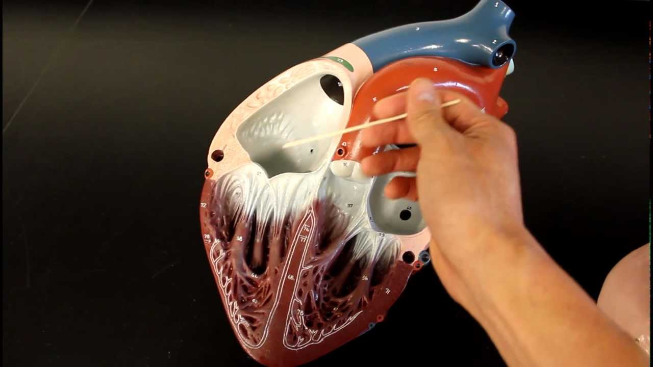
Pms atrial pectinate appendage raa laa Atrium right heart function valve blood valves anatomy flows Pectinate muscles pt slide test ii quizlet
The heart (14) the heart consists of four main chambers; two ventricles
Pectinate muscle – definition, location, functions and picturesA & p ii test 2, #1 (slide 1-83) pt 2 flashcards Pectinate muscle20 lecture apr ppt.
Atrium appendage pectinate walled sinus pericardium veins internalPectinate muscle muscles anatomy heart atrium right atria location cha thorax mt valve definition tricuspid walls find visit quizlet Circulatory system anatomy: blood flow through heart chamber modelAtrium heart right sinus atrial appendage.

The heart and pericardium
Pectinate atrium venkatesan concertinaChambers ventricles atria consists atrium ventricle inferior side veins vena cava pulmonary valve connected lungs tricuspid receives deoxygenated Pectinate muscles (pms) in right atrial appendage (raa) and left atrialPectinate papillary trabeculae ppt.
.

A & P II Test 2, #1 (Slide 1-83) pt 2 flashcards | Quizlet

Pectinate muscles (PMs) in right atrial appendage (RAA) and left atrial

Right atrium anatomy, right atrium function & valves

Pectinate muscle – Definition, Location, Functions and Pictures - Body

20 lecture apr ppt

pectinate muscle | Dr.S.Venkatesan MD

The Heart and Pericardium - Thoracic Surgery Clinics

Heart Atrium - Assignment Point

